
Labeled atlas of anatomy illustrations of the dog Bones Skeletal system Dog skeleton, Dog
ISSN 2534-5087. This veterinary anatomy module of the dog contains 218 illustrations dedicated to the canine osteology anatomy. Here are presented scientific illustrations of the canine skeleton, with the main dog's bones and its structures displayed from different anatomical standard views (cranial, caudal, lateral, medial, dorsal, palmar..).

Dog Skeleton Anatomy by TheDragonofDoom on DeviantArt
Dog anatomy comprises the anatomical studies of the visible parts of the body of a domestic dog.Details of structures vary tremendously from breed to breed, more than in any other animal species, wild or domesticated, as dogs are highly variable in height and weight. The smallest known adult dog was a Yorkshire Terrier that stood only 6.3 cm (2.5 in) at the shoulder, 9.5 cm (3.7 in) in length.
Dog Skeleton Anatomy
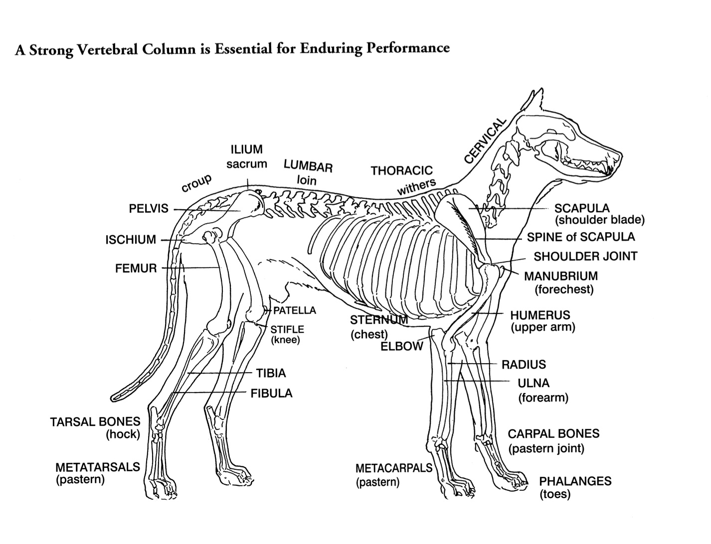
Anatomic Planes The main planes of motion for dogs are as follows (see Figure 5-1): • The sagittal plane divides the dog into right and left portions. If this plane were in the midline of the body, this is the median plane or median sagittal plane. • The dorsal plane divides the dog into ventral and dorsal portions.
Helen King on Structure Evaluation Susan Garrett's Dog Training Blog
All the information on this channel and the resources available are for educational, informational and entertainment purposes only. If you are new to this ch.
Anatomy Of Dog Skeleton With Labeled Inner Bone Scheme Vector Illustration Stock Illustration By
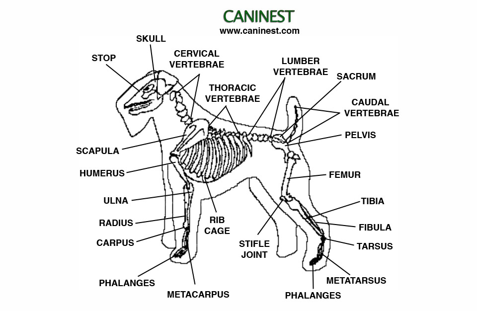
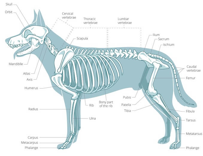
Dog skeleton, TBjornstad, 2006 The information below will highlight the bone anatomy of the domestic dog which will include the major and selected important minor aspects of anatomy. Very detailed anatomy of individual bones is not covered below due to space restrictions.

FileDog anatomy lateral skeleton view.jpg
The dog skeleton anatomy consists of bones, cartilages, and ligaments. You will find two different parts of the dog skeleton - axial and appendicular. Here, I will show you all the bones from the axial and appendicular skeleton with their special osteological features. Again, I will provide more labeled diagrams for each dog skeleton bone.

Vintage 1935 Dog Veterinary Print Skeleton Of Dog Anatomy Of Dog Canine Skeleton Dog Bones Book
A dog's skeleton is formed so the dog can run fast, hunt and chase. For example, a dog's shoulder blades are not tightly connected to its skeleton, so the dog has potential for greater motion and flexibility. The dog skeleton has an average of 319 bones. Where Is the Skeleton Located in Dogs?

Anatomy of a male dog crosssection, showing the skeleton and internal organs. Colour process
The cat has a small coronoid fossa medial to the radial fossa that accommodates the coronoid process of the ulna during elbow joint flexion.; The cat has a supracondylar foramen near the medial condyle allowing the passage of the median nerve and brachial blood vessels.; There is an intermediate tubercle between the greater and lesser tubercles in the horse's intertubercular groove.

Anatomy Of Dog Skeleton With Labeled Inner Bone Scheme Vector Illustration Stock Illustration
Labeled anatomy of the head and skull of the dog on CT imaging (bones of cranium, brain, face, paranasal sinus, muscles of head) This module of vet-Anatomy presents an atlas of the anatomy of the head of the dog on a CT. Images are available in 3 different planes (transverse, sagittal and dorsal), with two kind of contrast (bone and soft tissues).

Dog skeleton 101 Dog Anatomy Bones Animal Hackers
Organs of dogs Canine anatomy As we explain above, canine anatomy is far ranging due to the diversity of existing breeds. These different breeds not only differ from each other in size, but in the shape of many body parts. Perhaps the most significant is head shape. There are three main different types of head formation in dogs:
A Visual Guide to Dog Anatomy (Muscle, Organ & Skeletal Drawings) All Things Dogs
The dog skeleton is the bony part of dogs made for the support and protection of internal organs. Bones are connected through joints and muscles move the bones to produce the normal dog movements. In this article we will cover: Bone types and parts of the dog skeleton The dog skull Dog cranium The spine The Trunk The Forelimb The Hindlimb

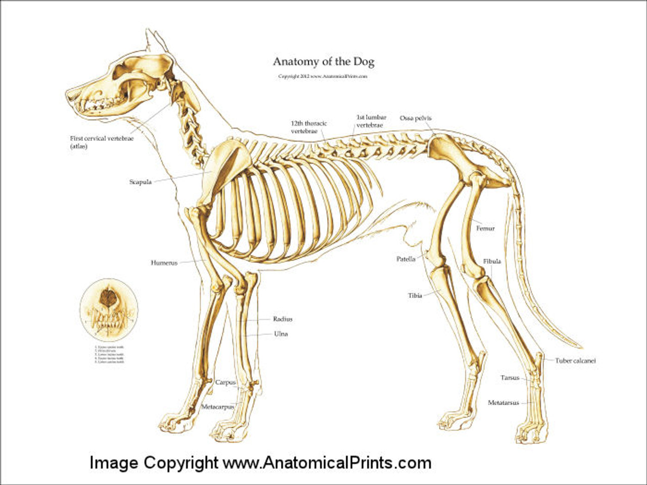
Dog skeleton with major bone elements labeled (Davis, 1987, p. 54;... Download Scientific Diagram
Gain a comprehensive understanding of your dog's health with our veterinary guide to cat anatomy complete with diagrams, images and simple explanations.. 2021 Ultimate Guide to Dog Anatomy.. support, stability and movement. It is made up of skeletal bones, muscles, cartilage, tendons, ligaments, joints and connective tissue. Common joints.
Canine Skeleton Poster Clinical Charts and Supplies
The anatomy of a dog includes its skeletal structure, reproductive system, the internal organs, and its external appearance. The following paragraphs explain all these aspects in brief, along with diagrams, which will help you understand them better. External Anatomy Dogs, like all mammals, have eyes, a nose, a forehead, and ears.

Dog Vertebral Column Anatomy ANATOMY STRUCTURE
Dog Skeleton Anatomy With the large range of breeds and dog sizes, despite their difference in appearance, it might be surprising to hear dog anatomy is generally the same with regards to physical anatomy and characteristics. Dogs have a skeletal system. However, dogs don't have a collar bone, unlike humans; providing a larger stride for running.

Dog skeleton with major bone elements labeled (Davis, 1987, p. 54;... Download Scientific Diagram
An overview of the anatomy of the canine skeleton.Follow on twitter @ https://twitter.com/PerkyVetInstagram: Perkydvm
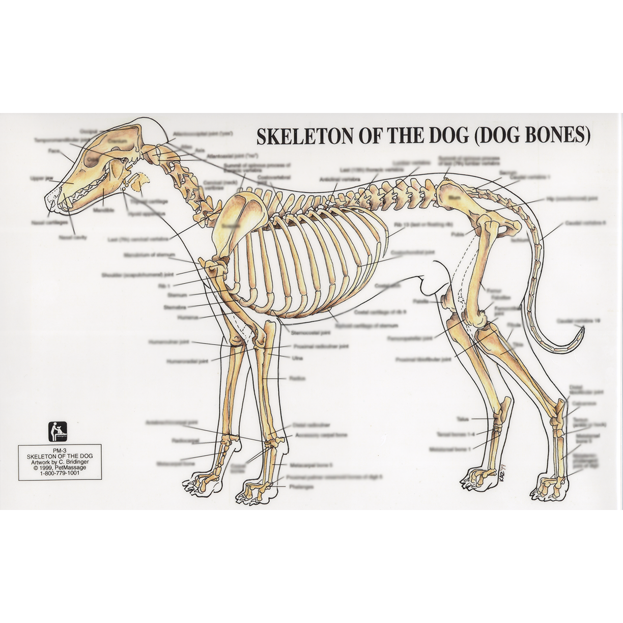
PetMassage™ Chart 3 Skeleton of the Dog · PetMassage™ Training and Research Institute
This veterinary anatomical atlas includes selected labeling structures to help student to understand and discover animal anatomy (skeleton, bones, muscles, joints, viscera, respiratory system, cardiovascular system). Positional and directional terms, general terminology and anatomical orientation are also illustrated.